Limited Edition Golden Llama is here! Check out how you can get one.
  Limited Edition Golden Llama is here! Check out how you can get one.
Offering SPR-BLI Services - Proteins provided for free! Offering SPR-BLI Services - Proteins provided for free!
Here come GMP Grade Cytokines!Free Sample is available! Here come GMP Grade Cytokines!Free Sample is available!
Click onto learn more what ACROBiosystems has developed to expedite your COVID-19 related research:
SARS-CoV-2 structures and life cycle (1)
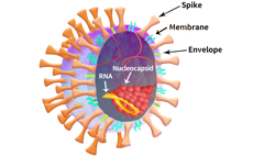
A: The virus particle structure of severe acute respiratory syndrome coronavirus 2 (SARS-CoV-2). The spherical particle of SARS-CoV-2 consists of four structural proteins: the spike proteins (S), the membrane protein (M), the envelope protein (E), and the nucleocapsid (N) protein. The S, M, and E proteins are incorporated in the virus membrane, while the N protein resides inside the particle and associated with virus genomic RNA.
B: Single-stranded positive-sense RNA genome of SARS-CoV-2. The ORF1a, or the ORF1a and ORF1b together, encodes the large polyproteins pp1a and pp1ab. The two polyproteins are further cleaved into 16 nonstructural proteins (nsp1–nsp16). The papain-like protease (PLpro), the 3C-like protease (3CLpro), the RNA-dependent RNA polymerase (RdRp), and the exonuclease (ExoN) are indicated. In addition to the nonstructural and structural proteins, the genome of SARS-CoV-2 also encodes six accessory proteins (Purple factors). Genome not drawn proportionally to size.
C: Structure of spike protein. The spike protein of SARS-CoV-2 is a trimer with each monomer comprising of an N-terminal S1 subunit and a C-terminal S2 subunit. The S1 subunit is further divided into the N-terminal domain (NTD), the receptor-binding domain (RBD), and C-terminal domain (CTD). During virus entry, the S1 subunit is responsible for receptor binding, while the S2 subunit is responsible for membrane fusion. Genome not drawn proportionally to size.
D: The life cycle of SARS CoV-2. Upon binding to the cellular receptor angiotensin-converting enzyme 2 (ACE2), the spike protein is activated by protease cleavage. Specifically, furin cleavage of S exposes a polyprotein basic sequence (RXXROH) on S1, a C-end rule (CendR) motif, that binds to cell surface Neuropilin-1 (NRP1) which may play a role in the increased infectivity of SARS-CoV-2. (2)
Additionally, based on diverse binding properties, biological functions, and clinical correlations analysis, Asialoglycoprotein receptor 1 (ASGR1) and Kringle domain-containing transmembrane protein 1 (KREMEN1) might be the alternative entry receptors which mediate virus entry independent of ACE2. (3)
After membrane fusion, the viral genomic RNA is released into the cytoplasm. The ORF1a and ORF1ab are translated into pp1a and pp1ab polypeptides, which are subsequently cleaved by PLpro and 3CLpro to produce nonstructural proteins (nsps). Several nsps, including nsps 7–10, nsp12, and nsp14, form the replication–transcription machinery to produce genomic and subgenomic RNAs. After the translation of structural proteins, the virion is assembled at the ER-Golgi intermediate compartment (ERGIC) and encapsulate the N protein and genomic RNA. The mature virion is released outside of cells by transportation through the vesicles. Virus replication at the steps of entry, proteolysis, and genome replication can be a therapeutic target for inhibition as indicated.
1. Ku Z. et al., Antibody Therapeutics, 2020, doi:10.1093/abt/tbaa007
2. Daly J. L. et al., Science, 2020, doi: 10.1126/science.abd3072
3. Gu Y. et al., biorxiv, 2020, doi: https://doi.org/10.1101/2020.09.09.287508


>>> Any requests, suggestions or feedback are welcome. click here.
This web search service is supported by Google Inc.


 A-Z
A-Z