Save Product

Product Details
Background
Recombinant Monoclonal Anti-PD-L1 Antibody, Rabbit (5D3) is a PD-L1 primary antibody for IHC application. PD-L1, is a ligand that binds with the receptor PD1, commonly found on T-cells, and acts to block T-cell activation. PD1 expression has been observed in a variety of cancers including melanoma and non-small cell lung cancer. The interaction of PD1/PDL1 is hypothesized to be a possible mechanism for the tumor to escape immune response. A number of checkpoint blockade inhibitors including pembrolizumab and nivolumab have been developed that target the PD1/PDL1 interaction in order to allow T-cells to recognize tumor cells without being deactivated by the tumor.
Product Specifications
Host SpeciesRabbitIsotypeRabbit IgG, Rabbit KappaClone5D3ApplicationIHC-PDilution Ratio1: 500 - 1: 2000FormLiquidClonalityMonoclonalReactivityHumanSynonymsCD274, PD-L1Concentration0.9~1.1 mg/mlSpecificityReact with PD-L1 protein on the membrane of target cell/tissuePurification TechProtein APreservationTris buffer, 1% BSAExperiment Protocol
Storage
Shipped at 4°C, Blue Ice. Store at +4°C for short term (1-2 weeks). Store at -20°C for long term.
Avoid freeze / thaw cycle. Upon delivery aliquot. The shelf life is 30 days from the date of opening.ACRO Quality Management System
Customers Also Viewed
Performance Data
Sample Staining Data
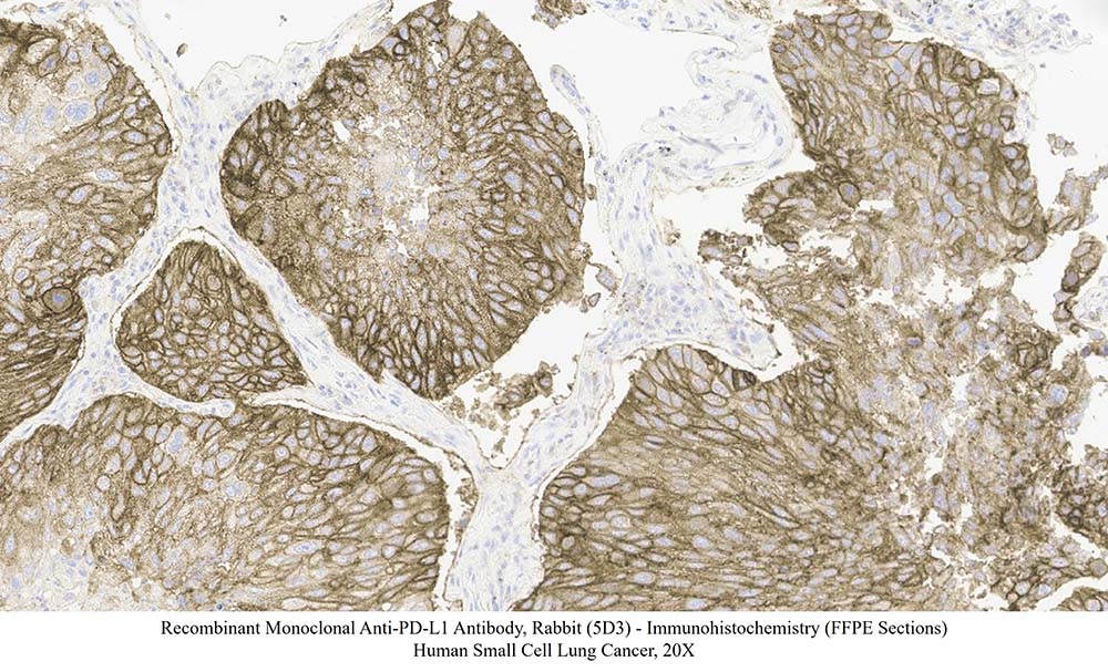
Immunohistochemical analysis of paraffin embedded human small cell lung cancer tissue labelled with HCS-S240 at 1/1000 dilution.
Perform heat mediated antigen retrieval with Tris/EDTA buffer pH 9.0 before commencing with LEICA BOND IHC staining protocol.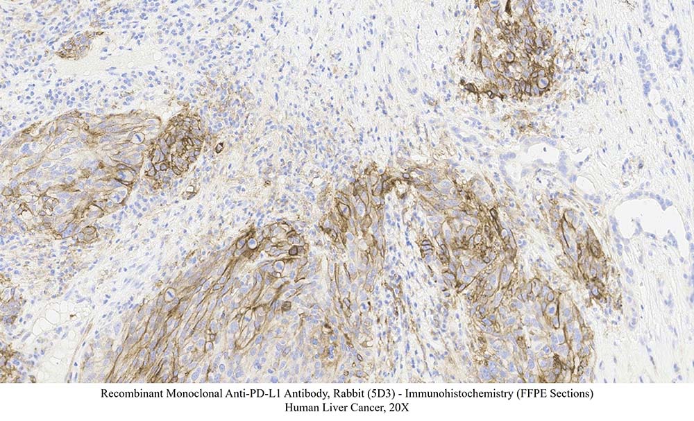
Immunohistochemical analysis of paraffin embedded human liver cancer tissue labelled with HCS-S240 at 1/1000 dilution.
Perform heat mediated antigen retrieval with Tris/EDTA buffer pH 9.0 before commencing with LEICA BOND IHC staining protocol.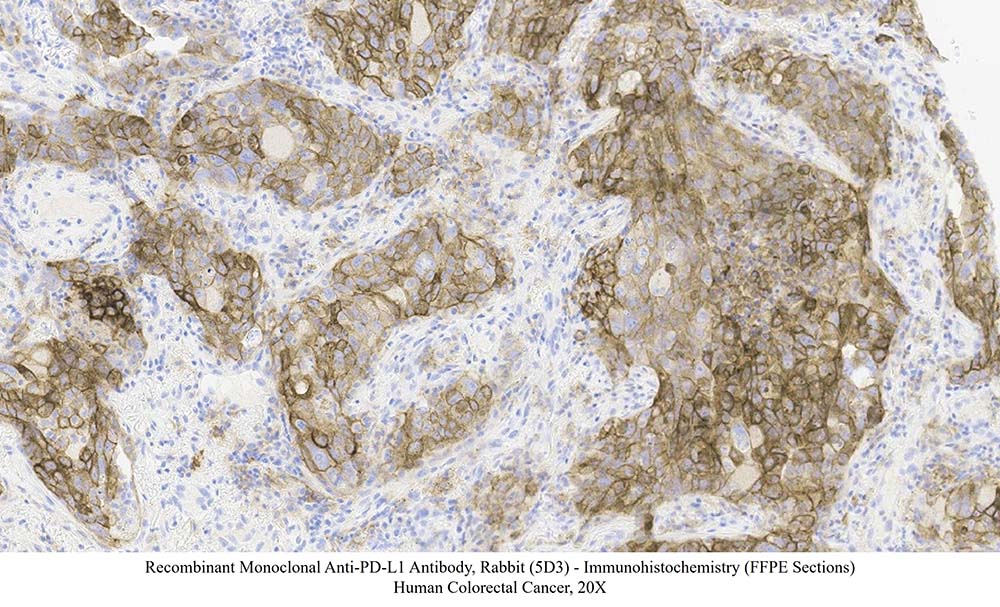
Immunohistochemical analysis of paraffin embedded human colorectal cancer tissue labelled with HCS-S240 at 1/1000 dilution.
Perform heat mediated antigen retrieval with Tris/EDTA buffer pH 9.0 before commencing with LEICA BOND IHC staining protocol.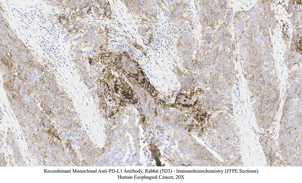
Immunohistochemical analysis of paraffin embedded human esophageal cancer tissue labelled with HCS-S240 at 1/1000 dilution.
Perform heat mediated antigen retrieval with Tris/EDTA buffer pH 9.0 before commencing with LEICA BOND IHC staining protocol.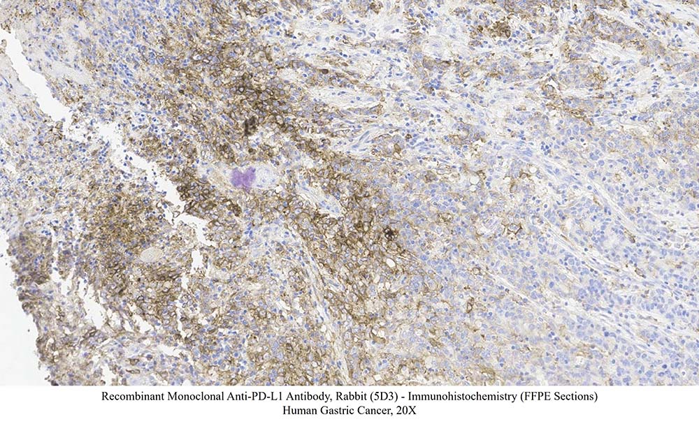
Immunohistochemical analysis of paraffin embedded human gastric cancer tissue labelled with HCS-S240 at 1/1000 dilution.
Perform heat mediated antigen retrieval with Tris/EDTA buffer pH 9.0 before commencing with LEICA BOND IHC staining protocol.
Customer Reviews Writing Reviews

Recent Advances
 Drug Development Progress
- English Name:
Programmed death-ligand 1
- Category:
- Approved Drugs:
12 Details
- Drugs in Clinical Trials:
176 Details
- Highest Development Stage:
Approved
 Contact Us

전화
글로벌: +1 800-810-0816 (무료)
아시아태평양: +86 400-682-2521
 Drug Candidate Licensing