Nuclease Activity Detection Kits

Deoxyribonucleases (DNases) and ribonucleases (RNases) are hydrolytic enzymes that degrade DNA and RNA by cleaving phosphodiester bonds. They are widely present in laboratory environments, raw materials, and consumables, and may also persist as process-related residues during biopharmaceutical manufacturing. Residual nucleases can compromise nucleic acid integrity, interfere with analytical workflows, and pose potential safety risks due to immunogenicity or unintended nucleic acid degradation. Therefore, sensitive and reliable detection of residual nuclease activity is critical throughout biopharmaceutical research, development, and production.
Conventional methods such as gel electrophoresis and UV spectrophotometry are commonly used to detect residual DNase and RNase activity, but they have limitations in sensitivity, quantification accuracy, throughput, or instrumentation requirements. In contrast, fluorescence probe–based assays offer high sensitivity, rapid detection, and precise quantitative measurement, making them a preferred approach for reliable residual nuclease activity detection.
Following rigorous methodological validation, ACROBiosystems has independently developed fluorescence probe–based DNase Activity Detection Kit and RNase Activity Detection Kit. These kits are designed to detect DNase and RNase contamination in laboratory environments, consumables, and raw materials, and for accurate quantification of residual nuclease activity in biopharmaceutical products, supporting effective process control and quality assurance.

Assay Principle

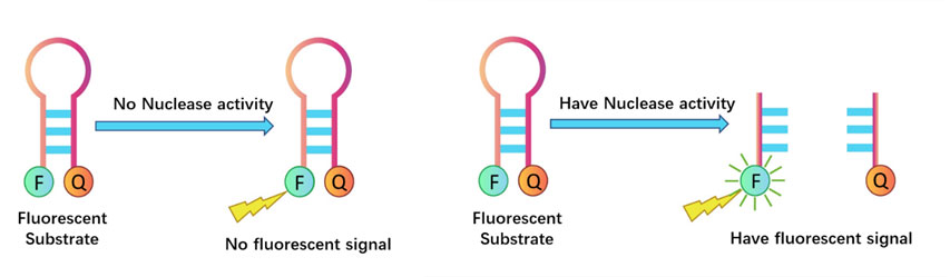
The fluorescence probe–based DNase and RNase activity assays utilize nucleic acid substrates labeled with fluorophore–quencher pairs. In the absence of DNase or RNase activity, the substrate remains intact, and the fluorophore is maintained in close proximity to the quencher. Due to fluorescence resonance energy transfer (FRET), fluorescence emission is effectively quenched, resulting in minimal background signal.When DNase or RNase activity is present, the nucleic acid substrate is cleaved, causing separation of the fluorophore and quencher. This disruption of the FRET effect leads to a progressively increasing fluorescence signal. The rate of fluorescence increase is directly proportional to the enzyme concentration and catalytic activity in the sample.
Fluorescence intensity is measured using a microplate reader at excitation/emission wavelengths of 490/520 nm for RNase and 535/565 nm for DNase. Based on the fluorescence signal, the presence of DNase or RNase contamination in the sample can be accurately determined.

Key Advantages

Based on enzymatic activity detection, suitable for residual DNase and RNase analysis.

Manufactured under stringent quality control to ensure high sensitivity, precision, accuracy, and stability.

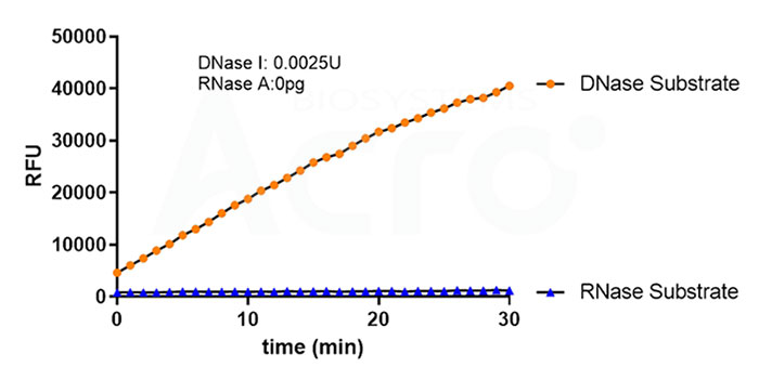
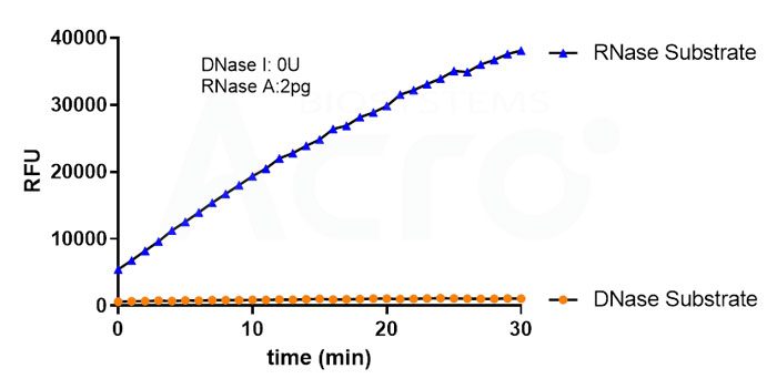
Enables parallel detection of DNase and RNase without cross-interference.

High sensitivity, with limits of detection as low as 3.9 × 10⁻⁵ U for DNase and 0.03 pg for RNase.

Simple and rapid workflow, allowing analysis of more than 80 samples within 30 minutes.

Assay Data
DNase Activity Assay Kit (Fluorescence)(Cat. No. ASE-A002
Interpretation Criteria
Microplate Reader Method: A fluorescence ratio (test sample/negative control) of < 2 indicates the absence of detectable DNase residue. A ratio ≥ 2 indicates the presence of DNase contamination or residual activity.
Application Scope
This kit has been validated for the detection of DNase contamination or residual activity in biologics, reagents, laboratory environments, and consumables. It is suitable for detecting a broad range of nucleases, including DNase I, Benzonase™, Exonuclease III, mung bean nuclease, micrococcal nuclease, Bal31 nuclease, S1 nuclease, and T7 endonuclease.
Product Performance
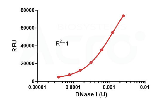
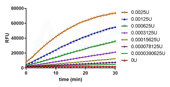
> High Sensitivity:Based on fluorescence signal analysis within a 30-minute reaction, the assay achieves a detection sensitivity as low as 3.90625 × 10⁻⁵ U.
High Sensitivity
> Rapid Turnaround with Robust Quantification:The assay can be completed within 30 minutes. The standard curve demonstrates excellent linearity (R² > 0.99), enabling accurate quantification of DNase concentration in test samples.
Rapid Turnaround with Robust Quantification
> High Precision:Intra-assay variability ≤ 10%; inter-assay variability ≤ 15%.

Intra-Assay Statistics:

Sample 1 2 3 4 5 6 7
Number of Replicate 8 8 8 8 8 8 8
Mean RFU 2703 4170 6804 11847 19904 32163 45024
Standard Deviation 30 87 182 340 538 455 713
Coefficient of Variation (%) 1.1 2.1 2.7 2.9 2.7 1.4 1.6

Inter-Assay Statistics:

Sample 1 2 3 4 5 6 7
Number of Replicate 8 8 8 8 8 8 8
Mean RFU 2987 4711 8021 13914 23835 37728 51370
Standard Deviation 291 504 1025 1879 3323 5016 6482
Coefficient of Variation (%) 9.8 10.7 12.8 13.5 13.9 13.3 12.6
> High Recovery Recovery rates consistently fall within 80%–120%.

System 55 µg/mL of Pyrophosphatase
(n=2)
20 µg/mL Thermostable Inorganic Pyrophosphatase (n=2) 10% of (50mM Tris-HCl and 50% Glycerol) (n=2) 1×Reaction Buffer (n=2)
Sample Conc. (U) Calculated Conc. (U) Ave % RE Calculated Conc. (U) Ave % RE Calculated Conc. (U) Ave % RE Calculated Conc. (U) Ave % RE
Sample 1 0.002 0.002132 107 0.00201 100 0.002013 101 0.002183 109
Sample 2 0.0005 0.000534 107 0.000491 98 0.000502 100 0.000565 113
Sample 3 0.0001 0.000110 110 0.000101 101 0.000105 105 0.000118 118
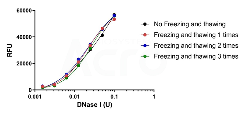
> Excellent Stability:After three freeze–thaw cycles, probe performance remains compliant with specifications, with no loss in sensitivity and a coefficient of variation (CV) < 10%.
RNase Activity Assay Kit (Fluorescence)(Cat. No. ASE-A001
Interpretation Criteria
Microplate Reader Method: A fluorescence ratio (test sample/negative control) of < 2 indicates the absence of detectable RNase residue. A ratio ≥ 2 indicates the presence of RNase contamination or residual activity.
Application Scope
This kit has been validated for the detection of RNase contamination or residual activity in biologics, reagents, laboratory environments, and consumables. It is suitable for detecting a broad range of nucleases, including RNase T1, RNase 1, micrococcal nuclease, Benzonase nuclease, mung bean nuclease, and S1 nuclease.
Product Performance
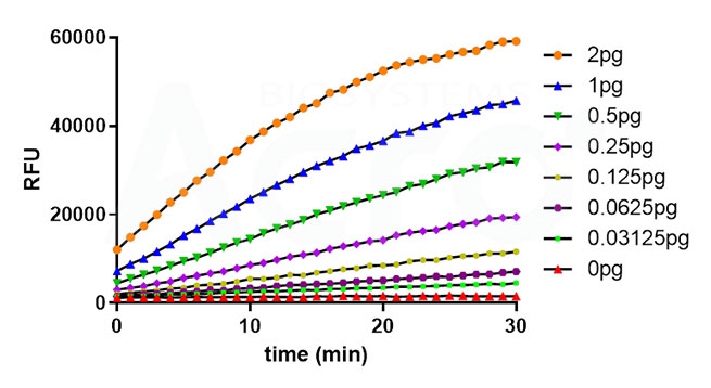
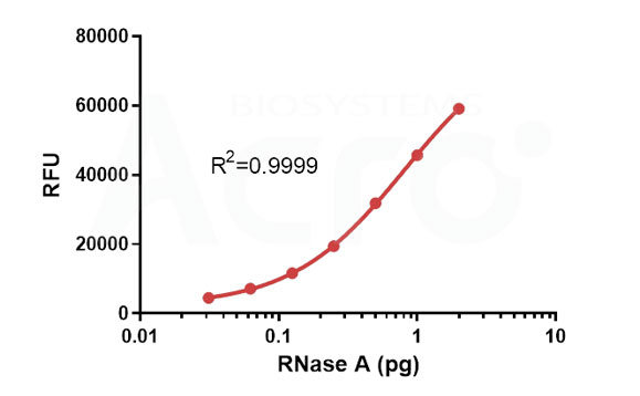
> High Sensitivity:Based on fluorescence signal analysis within a 30-minute reaction, the assay achieves a detection sensitivity as low as 0.03125 pg.
High Sensitivity
> Rapid Turnaround with Robust Quantification:The assay can be completed within 30 minutes. The standard curve demonstrates excellent linearity (R² > 0.99), enabling accurate quantification of RNase concentration in test samples.
•High Precision
> High Precision:Intra-assay variability ≤ 10%; inter-assay variability ≤ 15%.

Intra-Assay Statistics:

Sample 1 2 3 4 5 6 7
Number of Replicate 8 8 8 8 8 8 8
Mean RFU 3506 5964 9831 16862 27758 41779 56643
Standard Deviation 130 192 291 532 180 783 412
Coefficient of Variation (%) 3.7 3.4 3.0 3.2 0.6 1.9 0.7

Inter-Assay Statistics:

Sample 1 2 3 4 5 6 7
Number of Replicate 8 8 8 8 8 8 8
Mean RFU 3858 6132 10503 17609 28071 41161 53533
Standard Deviation 579 769 1495 2307 3341 4203 4249
Coefficient of Variation (%) 15 12.5 14.2 13.1 11.9 10.2 7.9
> High Recovery:Recovery rates consistently fall within 80%–120%.

System 55 µg/mL of Pyrophosphatase
(n=2)
20 µg/mL Thermostable Inorganic Pyrophosphatase (n=2) 10% of (50mM Tris-HCl and 50% Glycerol) (n=2) 1×Reaction Buffer (n=2)
Sample weight. (pg) Calculated weight. (pg) Ave % RE Calculated weight. (pg) Ave % RE Calculated weight. (pg) Ave % RE Calculated weight. (pg) Ave % RE
Sample 1 1.5 1.4252 95 1.3375 89 1.3319 89 1.4193 95
Sample 2 0.2 0.1691 85 0.1683 84 0.1669 83 0.1839 92
Sample 3 0.05 0.0437 87 0.0442 88 0.0461 92 0.0472 94
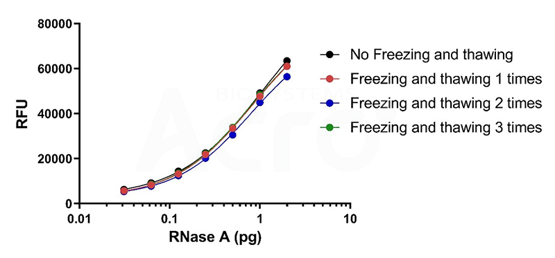
> Excellent Stability:After three freeze–thaw cycles, probe performance remains within specifications, with no reduction in sensitivity and a coefficient of variation (CV) < 10%.

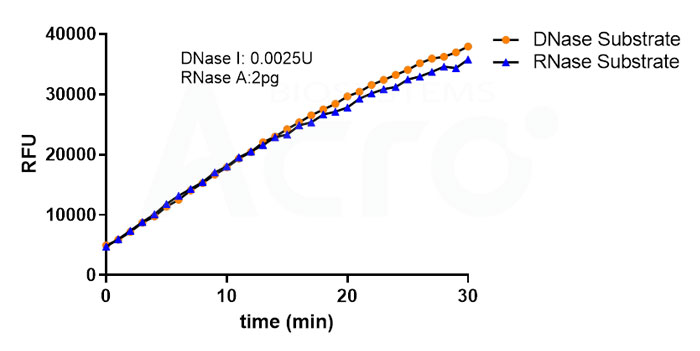
> No Cross-Interference:Validation studies confirm that when testing the same sample using the DNase Activity Assay Kit (Fluorescence) (Cat. No. ASE-A002) and the RNase Activity Assay Kit (Fluorescence) (Cat. No. ASE-A001), no cross-interference is observed.
No Cross-Interference
  • Background
  • Assay Principle
  • Key Advantages
  • Assay Data
Contact Us

전화
글로벌: +1 800-810-0816 (무료)
아시아태평양: +86 400-682-2521

주소 서울특별시 송파구 법원로11길 11, 제3층 제비-314,315호